Reperfusion Injury Risk Calculator
Calculate Reperfusion Injury Risk
When blood finally rushes back to tissue that’s been starved of oxygen, you’d think things get better right away. In reality, that sudden surge can cause a second wave of damage - a paradox that’s puzzled doctors for decades. This article unpacks why oxygen, the very molecule we breathe to stay alive, becomes a double‑edged sword during reperfusion injury.
What Is Reperfusion Injury?
Reperfusion injury is the tissue damage that occurs when blood supply returns to an organ after a period of ischemia (lack of oxygen). The abrupt restoration brings not only nutrients but also a flood of oxygen that can spark harmful chemical reactions.
Clinicians first noticed this phenomenon in heart attacks and strokes, where patients sometimes worsened after a blocked artery was reopened. Since then, researchers have linked reperfusion injury to organ transplants, traumatic injuries, and even surgical procedures that require temporary blood flow interruption.
Why Oxygen Becomes a Problem
Oxygen is essential for cellular metabolism, but when it re‑enters a deprived cell it can generate reactive oxygen species (ROS) - highly unstable molecules that attack DNA, proteins, and lipids.
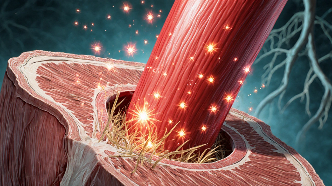
Think of ROS as mischievous sparks in a dry forest. A single spark can explode into a wildfire, and in a cell, those sparks quickly become a cascade of damage.
Key ROS generated during reperfusion include superoxide anion (O₂⁻), hydrogen peroxide (H₂O₂), and the hydroxyl radical (·OH). Each has distinct reactivity, but all share the ability to cripple cellular structures.
Cellular Targets of Oxygen‑Driven Damage
- Mitochondria: The powerhouses become leaky, releasing more ROS and triggering the opening of the mitochondrial permeability transition pore, which can lead to cell death.
- Lipid membranes: ROS attack phospholipids, producing lipid peroxides that disrupt membrane integrity and signaling.
- DNA: Oxidative lesions such as 8‑oxoguanine impair replication and can cause mutations.
- Proteins: Oxidation alters enzyme activity, especially those involved in calcium handling and contraction.
When these components fail together, the tissue experiences what researchers call the “no‑reflow” phenomenon, where micro‑vessels remain blocked despite the larger artery being open.
How the Body Reacts - The Inflammatory Cascade
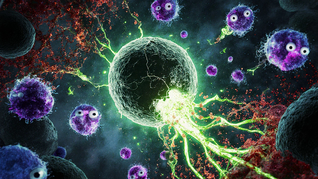
Oxygen‑induced ROS also act as alarm signals. They activate transcription factors like NF‑κB, which turn on genes for cytokines, chemokines, and adhesion molecules. The result? Neutrophils and other immune cells swarm the area, releasing more ROS, proteases, and inflammatory mediators.
One striking example is the release of myeloperoxidase from neutrophils, which converts H₂O₂ into the even more potent hypochlorous acid (HOCl). This amplifies the oxidative assault and extends damage beyond the originally ischemic zone.
Protective Roles of Oxygen - When It Helps
It’s not all doom and gloom. Controlled oxygen delivery can trigger protective pathways. Pre‑conditioning - brief, non‑lethal episodes of ischemia followed by reperfusion - teaches the heart to cope with oxidative stress. During these episodes, a modest rise in ROS activates antioxidant enzymes such as superoxide dismutase (SOD) and catalase, bolstering the tissue’s defenses.
Additionally, nitric oxide (NO) produced by endothelial cells can scavenge superoxide, forming peroxynitrite (ONOO⁻). While peroxynitrite is itself reactive, low‑level formation can modulate vascular tone and improve micro‑circulation.
Clinical Strategies to Tame Oxygen’s Dark Side
Doctors and researchers have tried several approaches to limit oxidative injury while still restoring blood flow.
- Antioxidant Therapy: Agents like N‑acetylcysteine (NAC) and vitamin C act as scavengers for ROS. Clinical trials in myocardial infarction show modest reductions in infarct size when given early.
- Ischemic Post‑Conditioning: Brief, intermittent interruptions of blood flow right after reperfusion can reduce ROS spikes and improve outcomes.
- Targeted Enzyme Inhibitors: Drugs that inhibit xanthine oxidase (e.g., allopurinol) lower superoxide production during re‑oxygenation.
- Controlled Oxygen Delivery: Using lower FiO₂ (fraction of inspired oxygen) levels during resuscitation avoids hyperoxia, which would otherwise overwhelm antioxidant systems.
- Mitochondrial Protective Agents: Compounds like cyclosporine A block the mitochondrial permeability transition pore, preserving cellular energy.
Each method aims to balance the need for oxygen with the risk of oxidative overload.
Key Differences: Early vs. Late Phase Effects
| Phase | Dominant ROS | Cellular Impact | Therapeutic Focus |
|---|---|---|---|
| Early | Superoxide, Hydrogen peroxide | Mitochial permeability, lipid peroxidation, Ca²⁺ overload | Rapid antioxidant infusion, post‑conditioning |
| Late | Hydroxyl radical, Peroxynitrite | Inflammatory cell infiltration, extracellular matrix degradation | Anti‑inflammatory agents, nitric oxide modulation |
Practical Take‑aways for Clinicians and Researchers
- Monitor oxygen levels closely during resuscitation; avoid hyperoxia.
- Consider early antioxidant administration in high‑risk reperfusion scenarios.
- Use ischemic post‑conditioning protocols where feasible - a few seconds of clamping can make a big difference.
- Stay aware of patient‑specific factors like diabetes or chronic inflammation, which can amplify oxidative stress.
- Follow emerging research on mitochondria‑targeted drugs; they may become standard of care in the next few years.
Understanding the dual nature of oxygen helps clinicians turn a potential bug into a feature of recovery.
Frequently Asked Questions
What exactly causes the damage when oxygen returns to tissue?
The surge of oxygen creates reactive oxygen species that attack cell membranes, DNA, and proteins. The resulting oxidative stress triggers inflammation and can open mitochondrial pores, leading to cell death.
Is giving patients extra oxygen always harmful?
Not necessarily. Controlled oxygen levels are essential to prevent hypoxia. The danger lies in hyperoxia - too much oxygen too quickly - which overwhelms the body’s antioxidant defenses.
How do antioxidants help during reperfusion?
Antioxidants neutralize ROS before they can damage cellular components. For example, N‑acetylcysteine donates cysteine to replenish glutathione, a key intracellular scavenger.
Can lifestyle changes reduce the risk of reperfusion injury?
Yes. Regular aerobic exercise boosts endogenous antioxidant enzymes, and a diet rich in polyphenols (berries, nuts) provides additional ROS‑scavenging compounds.
What future therapies are being explored?
Researchers are testing mitochondria‑targeted peptides, gene‑editing approaches to up‑regulate antioxidant enzymes, and nanocarriers that deliver antioxidants directly to ischemic tissue.
11 Comments
One cannot help but notice the ubiquitous yet unspoken agendas that pervade contemporary biomedical discourse, wherein authors subtly obfuscate the true ramifications of oxidative reperfusion. The prevailing dogma, replete with half‑trite metaphors of “sparks” and “wildfires,” merely masks a deeper industrial collusion to perpetuate pharmaceutical dependence. Moreover, the cursory acknowledgment of ROS as a singular villain betrays a lazily curated narrative that eschews the nuanced interplay of redox biology.
Indeed, the biochemical cascade you describe is a marvel of nature-yet, it also demands our vigilance, our compassion, and, above all, our rigorous scientific scrutiny, for only through such balanced inquiry can we hope to mitigate the deleterious sequelae of reperfusion injury, and perhaps pave the way for therapeutic breakthroughs, without succumbing to dogmatic inertia.
It’s heartbreaking to think patients might suffer more after the very treatment meant to save them.
Allow me to clarify the mechanistic underpinnings with unequivocal precision: upon re‑oxygenation, electron leakage from complex I and III of the mitochondrial electron transport chain precipitates the rapid formation of superoxide anion, which is subsequently dismutated to hydrogen peroxide; in the presence of transition metals, the Fenton reaction yields the hydroxyl radical, the most indiscriminate oxidant of cellular macromolecules. This sequence, corroborated by numerous spectroscopic studies, underscores the inevitability of oxidative stress in the immediate post‑ischemic window. 😊
Yo, the whole NADH‑driven ROS surge is basically a bio‑feedback loop that’s dummified by hyperoxic protocols, ya feel? If we don’t modulate FiO2, we just amplify the redox load, leading to what the lit calls “no‑reflow” phenomena.
Remember, every clinician holds the power to turn this double‑edged sword into a shield; by embracing controlled oxygen titration and early antioxidant infusion, we can chart a course toward resilience. Let us rally behind evidence‑based protocols, championing the lives of those who entrust us with their recovery. Together, we shall rewrite the narrative of reperfusion injury.
Man, it’s like the shadowy cabal of big pharma wants us to keep pumping high‑flow oxygen just to sell more meds, while the real fix sits hidden in low‑dose strategies they refuse to publicize.
It is a moral imperative for the medical community to confront the paradoxical nature of oxygen therapy with unflinching honesty, for time and again we have witnessed the tragic consequences of neglecting the very principles we claim to uphold. When a vessel is finally reopened after a protracted occlusion, the sudden influx of O₂, though essential for cellular respiration, initiates a cascade of oxidative reactions that betray the intended healing process. This betrayal is not a mere biochemical curiosity; it translates into palpable morbidity, extended hospital stays, and, in the gravest cases, preventable mortality. The early phase, dominated by superoxide and hydrogen peroxide, indiscriminately assaults mitochondrial membranes, prompting the opening of the permeability transition pore and precipitating necrotic cell death. In the later phase, the relentless onslaught of hydroxyl radicals and peroxynitrite fuels an inflammatory maelstrom, recruiting neutrophils that exacerbate tissue destruction through proteolytic enzymes and additional ROS. Yet, despite this wealth of mechanistic insight, clinical practice persistently clings to antiquated protocols that prioritize rapid reperfusion without due consideration of oxidative moderation. It is incumbent upon us to champion evidence‑based interventions such as ischemic post‑conditioning, judicious oxygen titration, and targeted antioxidant therapy, lest we remain complicit in a cycle of iatrogenic harm. Moreover, the ethical dimension extends beyond individual patient outcomes; the systemic overuse of high‑flow oxygen imposes a fiscal burden on healthcare systems already strained by escalating costs. By embracing a philosophy that balances oxygen's life‑sustaining virtues against its capacity for destruction, we honor the Hippocratic oath in its fullest sense. Let us, therefore, demand rigorous training, continuous monitoring, and interdisciplinary collaboration to refine our approach to reperfusion. In doing so, we not only mitigate oxidative injury but also reaffirm our commitment to the sanctity of human life, transcending the mere technicalities of pharmacology and physiology. The path forward is clear: measured, compassionate, and scientifically grounded care must supplant reflexive hyperoxia. We must also invest in translational research that brings mitochondrial protective agents from bench to bedside, ensuring that promising compounds such as cyclosporine A are evaluated in robust clinical trials. Education of frontline staff regarding the perils of hyperoxia should become a mandatory component of residency curricula, fostering a generation of physicians attuned to oxidative balance. Ultimately, the collective stewardship of oxygen will define the future of reperfusion therapy, and we cannot afford complacency.
From a global health perspective, it is essential to recognize that resource‑limited settings may lack access to sophisticated antioxidant regimens, making judicious oxygen use even more critical.
We should share low‑cost protocols worldwide and train staff to monitor oxygen saturation without excess devices
The battlefield of reperfusion demands both sword and shield, and we must wield them wisely.
Write a comment