
Imagine you are sitting on the couch, watching TV or reading a book, when suddenly it feels like someone has squeezed your chest. You try to take a deep breath, but the air just won't come in. Your heart starts racing. This isn't asthma. It isn't anxiety. For thousands of people every year, this is the terrifying first sign of a Pulmonary Embolism, also known as PE. It is a life-threatening blockage in one of the pulmonary arteries in your lungs caused by a blood clot that usually travels from your legs. The scariest part about a PE is how easily it gets missed. Because the symptoms mimic other common conditions, many patients wait days-or even weeks-for a correct diagnosis. In fact, data shows that nearly 70% of PE patients visit a healthcare provider multiple times before getting the right answer. Understanding what to look for and how doctors diagnose this condition can literally save your life.
The Hidden Danger: How a Clot Reaches Your Lungs
To understand why a PE happens so fast, you have to look at where the problem starts. Most of the time, the trouble begins in your legs. A blood clot forms in a deep vein, a condition called Deep Vein Thrombosis (or DVT). It is a blood clot that forms in a deep vein, most commonly in the lower leg, thigh, or pelvis. About 70% of all pulmonary embolisms originate from these leg clots. Here is what happens next: A piece of that clot breaks loose. It travels through your bloodstream, heading toward your heart. Once it reaches the right side of your heart, it gets pumped into the lungs. There, it lodges in the Pulmonary Arteries. These are the blood vessels that carry deoxygenated blood from the heart to the lungs for oxygenation. The artery is blocked. Blood can’t get through to pick up oxygen. Your body screams for air because it’s starving for it. This doesn’t happen overnight. Risk factors include long periods of immobility (like long flights or hospital stays), recent surgery, cancer, smoking, and certain genetic conditions. If you have had a DVT or PE before, your risk of recurrence jumps significantly-about one-third of patients will experience another event within ten years.
Sudden Shortness of Breath: The Cardinal Symptom
If there is one symptom that defines a pulmonary embolism, it is sudden shortness of breath. Medical reviews show this occurs in 85% of cases. But "shortness of breath" can mean different things. In a massive PE, the dyspnea is abrupt and profound. You might feel winded even while resting. In smaller, peripheral PEs, the shortness of breath might be mild or come and go, which makes it easy to dismiss as fatigue or stress. However, if you notice that your breathing becomes difficult without any physical exertion, pay attention. Other symptoms often accompany the breathlessness:
- Chest Pain: About 74% of patients experience chest pain. It is often sharp and worsens when you take a deep breath, cough, or bend over. This is called pleuritic pain. Many people mistake this for a heart attack, but the nature of the pain-sharp and linked to breathing-is a key clue.
- Rapid Heart Rate: Your heart tries to compensate for the lack of oxygen by beating faster. Tachycardia (heart rate over 100 bpm) is present in about 30% of cases.
- Coughing: Roughly half of patients cough. In about 23% of cases, they cough up small amounts of blood (hemoptysis).
- Fainting: Syncope occurs in 14% of cases, usually indicating a large clot burden and low blood pressure.
- Leg Swelling: Since most PEs start with a DVT, one leg may be swollen, red, or warm to the touch compared to the other.
The Diagnostic Puzzle: Scoring and Blood Tests
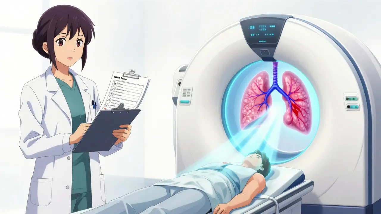
Diagnosing a PE is not as simple as running one test. Doctors use a step-by-step approach to avoid unnecessary radiation and contrast dye exposure while ensuring no dangerous clots are missed. The process usually starts with clinical probability scoring. The two most common tools are the Wells Criteria. This is a clinical prediction rule used to estimate the pre-test probability of pulmonary embolism based on patient history and physical exam findings. and the Geneva Score. This is another validated clinical decision rule for assessing the likelihood of pulmonary embolism, particularly useful in emergency settings. These scores assign points for symptoms like rapid heart rate, recent surgery, active cancer, and signs of DVT. They help doctors categorize you as low, moderate, or high risk. If your score suggests a low probability, the next step is usually a D-dimer Test. This is a blood test that measures fibrin degradation products, indicating whether a blood clot is breaking down in the body. D-dimer is a protein fragment released when a clot dissolves. A negative result (below 500 ng/mL) effectively rules out PE in low-risk patients with 97% sensitivity. However, D-dimer has a major flaw: it lacks specificity. It can be elevated due to infection, inflammation, pregnancy, or simply aging. In patients over 50, the accuracy drops significantly. That is why new guidelines recommend age-adjusted thresholds (e.g., adding 10 ng/mL for every year over 50). If your D-dimer is positive, or if your initial risk score was high, you move straight to imaging.
Imaging Gold Standards: CTPA and V/Q Scans
When blood tests aren't enough, imaging takes center stage. The gold standard for diagnosing PE is CT Pulmonary Angiography (or CTPA). It is a specialized CT scan using intravenous contrast dye to visualize blood flow in the pulmonary arteries and detect clots. During a CTPA, you receive an injection of iodinated contrast dye. The scanner takes detailed cross-sectional images of your lungs. It has a sensitivity of 95% and specificity of 96%, meaning it catches almost all significant clots. It provides a clear picture of where the clot is and how large it is. However, it involves radiation exposure (about 5-7 mSv) and requires your kidneys to process the contrast dye. For patients who cannot tolerate contrast dye-due to kidney failure or severe allergies-doctors use a Ventilation/Perfusion Scan (or V/Q Scan). It is a nuclear medicine test that compares airflow and blood flow in the lungs to identify mismatches indicative of a pulmonary embolism. You inhale a radioactive gas and receive an injection of a radioactive tracer. If air flows into a part of the lung but blood does not, it suggests a blockage. While slightly less sensitive than CTPA (85%), it avoids radiation to the abdomen and contrast risks. Another crucial tool is Compression Ultrasound. This is a non-invasive imaging technique using sound waves to detect blood clots in the deep veins of the legs. If you have leg swelling, doctors will scan your legs. Finding a DVT in a patient with respiratory symptoms is often treated as a PE, even if the lung scan is inconclusive, because the treatment is similar.
| Diagnostic Tool | Sensitivity | Specificity | Best Used When | Key Limitation |
|---|---|---|---|---|
| D-dimer Blood Test | 97% (Low Risk) | Variable (Low in elderly) | Ruling out PE in low-probability patients | False positives from inflammation/age |
| CTPA | 95% | 96% | First-line imaging for stable patients | Radiation exposure; contrast allergy/kidney issues |
| V/Q Scan | 85% | 95% | Patients with contrast contraindications | Less accurate if existing lung disease (COPD) |
| Compression Ultrasound | >90% (for DVT) | 95% | Patients with leg swelling/pain | Only detects clots in legs, not lungs directly |

Why Diagnosis Gets Delayed: Real-World Challenges
Despite advanced technology, misdiagnosis remains a huge problem. Why? Because PE is a "great mimicker." Its symptoms overlap with pneumonia, asthma, heart attacks, and even panic attacks. Patient stories highlight this frustration. Many report being told their shortness of breath was "just anxiety" or "deconditioning" after weeks of suffering. One survey found that 68% of PE patients saw a doctor an average of 2.3 times before the correct diagnosis was made. Initial misdiagnoses often included pneumonia or asthma. Several factors contribute to these delays:
- Non-Specific Symptoms: Mild shortness of breath and a dry cough don't always scream "emergency" to a tired ER doctor.
- Normal Chest X-Rays: Since PEs don't show up on standard X-rays, doctors might assume the lungs are healthy.
- Provider Bias: Younger patients, especially women, are sometimes dismissed as having anxiety-related hyperventilation.
- Lack of Clinical Suspicion: Without asking about recent travel, surgery, or family history of clots, doctors may not calculate a Wells Score.
What You Can Do: Advocacy and Awareness
If you experience sudden, unexplained shortness of breath, do not ignore it. Here is how to advocate for yourself: 1. Be Specific About Onset: Tell the doctor exactly when the symptoms started. Did it happen suddenly while resting? Did it get worse with deep breaths? 2. Mention Risk Factors: Bring up any recent long-distance travel, surgery, hospitalization, or history of clots. If you have cancer or are on hormone therapy, say so. 3. Ask About Probability Scores: You can politely ask, "Have we calculated my Wells Score or Geneva Score to assess my risk?" This prompts the clinician to follow the structured guideline. 4. Push for Imaging if Needed: If your D-dimer is positive or your risk is moderate/high, insist on understanding the plan for imaging. Don't settle for "wait and see" if you feel unstable. 5. Know the Emergency Signs: If you faint, have severe chest pain, or cough up blood, call emergency services immediately. Do not drive yourself.
Pulmonary embolism is serious, but it is treatable. Anticoagulants (blood thinners) are the standard treatment to prevent further clots. In severe cases, procedures to break up or remove the clot may be necessary. The key is early recognition. By understanding the symptoms and the diagnostic process, you empower yourself to seek the right care at the right time.
How quickly does a pulmonary embolism develop?
The symptoms of a PE typically appear suddenly. While the underlying clot (DVT) may have formed in the legs over days or weeks, the moment it breaks off and travels to the lungs, the onset of shortness of breath and chest pain is often abrupt and immediate.
Can a PE be mistaken for anxiety?
Yes, frequently. Both conditions cause rapid breathing, chest tightness, and a racing heart. However, anxiety rarely causes unilateral leg swelling or hemoptysis (coughing blood). If standard anxiety treatments don't resolve the symptoms, medical evaluation for PE is crucial.
Is a normal D-dimer test enough to rule out PE?
Only if your clinical probability score (Wells or Geneva) is low. If you have high-risk features like recent surgery, active cancer, or signs of DVT, a normal D-dimer might still require imaging because the test's reliability decreases in complex cases.
What is the survival rate for pulmonary embolism?
With timely diagnosis and treatment, the prognosis is good. Mortality rates have dropped from 8.5% in 2015 to around 5.2% recently due to better diagnostic protocols. However, untreated massive PE can be fatal within hours.
Who is at highest risk for developing a PE?
High-risk groups include individuals with active cancer, those who have recently undergone major surgery or trauma, people with prolonged immobility (bed rest or long flights), smokers, pregnant women, and those with a personal or family history of blood clots.
